Home » Without Label » Smooth Muscle Diagram - I Answer The Following A Draw A Labeled Diagram Of Smooth Muscle Give One Difference Between Yudem And Nh - Smooth muscle is found in the walls of hollow organs like your intestines and stomach.
Smooth Muscle Diagram - I Answer The Following A Draw A Labeled Diagram Of Smooth Muscle Give One Difference Between Yudem And Nh - Smooth muscle is found in the walls of hollow organs like your intestines and stomach.
Smooth Muscle Diagram - I Answer The Following A Draw A Labeled Diagram Of Smooth Muscle Give One Difference Between Yudem And Nh - Smooth muscle is found in the walls of hollow organs like your intestines and stomach.. Smooth muscles are unique in their largely involuntary response, and in their structure. Smooth muscle is a type of muscle tissue which is used by various systems to apply pressure to vessels and organs. Human muscles · august 4, 2020. • smooth muscles respond to stretch only briefly, and then adapts to its new length • the new length however, retains its original _____ seconds or minutes after it has been elongated or shortened (e.g. Smooth muscle contracts under certain stimuli as atp is freed.
It is layered in a distinctive pattern of circular layers. This page describes smooth muscle development, descriptions of cardiac muscle and smooth muscle development can be found in other notes. Its wavelike movements propel things through the bodily system, such as food through. It is the pen diagram of skeletal, smooth and cardiac muscle for class 10, 11 and 12. Smooth muscle anatomy smooth muscle tissue is also known as visceral muscle tissue.
Involuntary Muscle Definition And Examples Biology Online Dictionary from nitrocdn.com Smooth muscle fibers are often found forming sheets of tissue and function in a coordinated fashion due to the presence of gap junctions between the cells. Human muscles · august 4, 2020. Start studying smooth muscle tissue. Smooth muscle anatomy smooth muscle tissue is also known as visceral muscle tissue. This diagram shows a few of the cells that can be seen in the stained section below. During exercise, the smooth muscle in the blood vessels can restrict or increase blood flow through the blood vessel so that more blood carrying oxygen can go to the skeletal muscle. Smooth muscle cells lack the striated banding pattern found in cardiac and skeletal muscle, and they receive neural innervation from the autonomic nervous system. Learn vocabulary, terms, and more with flashcards, games, and other study tools.
This smooth muscle can be found surrounding the walls of the blood vessels, the bronchioles in the lungs, and the sphincter muscles used in the gi tract.the gi tract, which is tubular by design, also houses longitudinal muscles in addition to the smooth.
Smooth muscle is widely distributed in the body. Note that the smooth muscle cells are arranged in layers that are orthagonal to each other. Start studying smooth muscle tissue. Smooth muscle is made up of cells that contain a single central nucleus. Smooth muscles are unique in their largely involuntary response, and in their structure. It constitutes much of the musculature of This page describes smooth muscle development, descriptions of cardiac muscle and smooth muscle development can be found in other notes. Human muscles · august 4, 2020. Gap junctions between cells allows coordination of contraction. Smooth muscle is unconsciously controlled by the nervous system; Smooth muscle is a type of muscle tissue which is used by various systems to apply pressure to vessels and organs. Learn vocabulary, terms, and more with flashcards, games, and other study tools. Smooth muscle often contracts an organ in multiple.
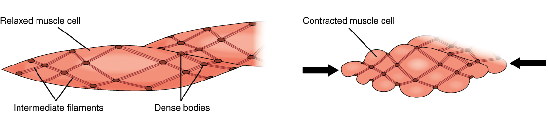
• smooth muscles respond to stretch only briefly, and then adapts to its new length. In this video i have shown the simplest way of drawing muscle drawing. Note that the smooth muscle cells are arranged in layers that are orthagonal to each other. Smooth muscle makes up the walls of hollow organs, respiratory passageways, and blood vessels. Smooth muscle tissue, unlike striated muscle, contracts slowly and automatically.
Smooth Muscle Anatomy And Physiology from opentextbc.ca Smooth muscle is widely distributed in the body. Distinguish between skeletal muscle and smooth muscle. Smooth muscle cells are under control of the autonomous nervous system. In this video i am gonna to show you how to draw the diagrams of cardiac, straited, smooth muscle for class 1st to 10th. The cells are spindle shaped, and the nucleus is central. The cells stick together and are connected by specialised cell junctions, called gap junctions. Smooth muscle tissue, unlike striated muscle, contracts slowly and automatically. Smooth muscle anatomy smooth muscle tissue is also known as visceral muscle tissue.
In skeletal muscle, a single type of somatic nervous system traverses to muscle, where it stimulates organelle in the muscle cells in order to release calcium. Smooth muscle determines the flow of blood in the arteries. Smooth muscle is made up of cells that contain a single central nucleus. The smooth muscles perform the functions in the contrast of other types of muscles. Note that the smooth muscle cells are arranged in layers that are orthagonal to each other. Related posts of smooth muscle labelled diagram muscle anatomy lower extremity. Smooth muscle contracts under certain stimuli as atp is freed. • smooth muscles respond to stretch only briefly, and then adapts to its new length • the new length however, retains its original _____ seconds or minutes after it has been elongated or shortened (e.g. Smooth muscle is found in the walls of hollow organs like your intestines and stomach. The cells are spindle shaped, and the nucleus is central. Draw the diagram of a sarcomere of skeletal muscle showing different regions. Therefore, performers get the oxygen their muscles need whilst exercising Smooth muscle fibers are often found forming sheets of tissue and function in a coordinated fashion due to the presence of gap junctions between the cells.
It is layered in a distinctive pattern of circular layers. In arteries, smooth muscle movements maintain the arteries' diameter. Note that the smooth muscle cells are arranged in layers that are orthagonal to each other. It constitutes much of the musculature of Smooth muscles in arteries and veins are largely responsible for regulation of blood pressure.
Histology Slides Database Smooth Muscle High Resolution Histology Diagram from lh6.ggpht.com Smooth muscles are unique in their largely involuntary response, and in their structure. It also occurs in the spleen (capsule and trabeculae), eye (iris and ciliary body), skin (arrector pili muscles of hairs. Smooth muscle (textus muscularis levis) smooth muscle is a type of tissue found in the walls of hollow organs, such as the intestines, uterus and stomach. • smooth muscles respond to stretch only briefly, and then adapts to its new length • the new length however, retains its original _____ seconds or minutes after it has been elongated or shortened (e.g. • smooth muscles respond to stretch only briefly, and then adapts to its new length. Note that the smooth muscle cells are arranged in layers that are orthagonal to each other. In this video i have shown the simplest way of drawing muscle drawing. The cells stick together and are connected by specialised cell junctions, called gap junctions.
Smooth muscles are structurally the simplest of all muscles types.
Smooth muscles are structurally the simplest of all muscles types. Smooth muscle fibers are often found forming sheets of tissue and function in a coordinated fashion due to the presence of gap junctions between the cells. The cells stick together and are connected by specialised cell junctions, called gap junctions. Smooth muscle makes up the walls of hollow organs, respiratory passageways, and blood vessels. The muscle tissue (patrick steele) Smooth muscle is widely distributed in the body. Smooth muscle determines the flow of blood in the arteries. • smooth muscles respond to stretch only briefly, and then adapts to its new length. Smooth muscles in arteries and veins are largely responsible for regulation of blood pressure. It is layered in a distinctive pattern of circular layers. Muscle anatomy lower extremity 12 photos of the muscle anatomy lower extremity anatomy lower extremity muscle quiz, lower extremity muscle anatomy ct, lower extremity muscle anatomy mri, muscle anatomy lower extremity, muscular anatomy of lower limb, human muscles, anatomy lower extremity muscle quiz, lower. Arteries have thick walls due to smooth muscle cells, which help them carry blood away from the heart to every part of. Smooth muscles are unique in their largely involuntary response, and in their structure.